Importance of Focus in OCT Angiography
Alexander Tomlinson, Bilal Hasan, and Brandon J. Lujan
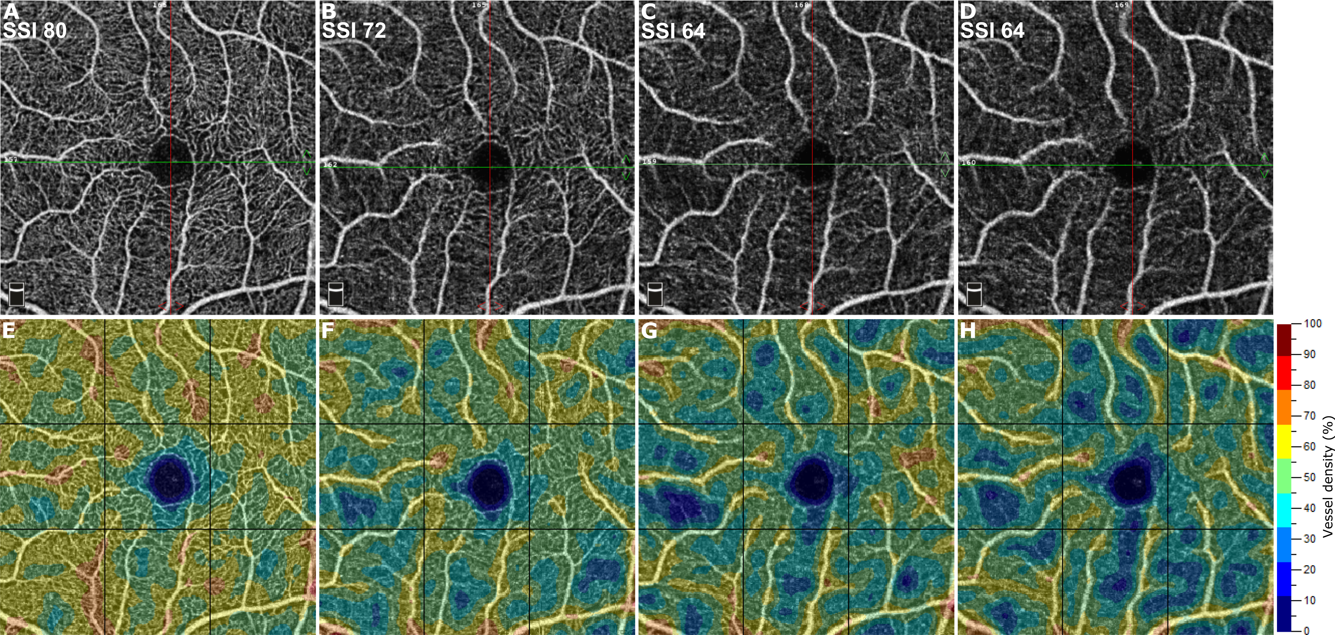
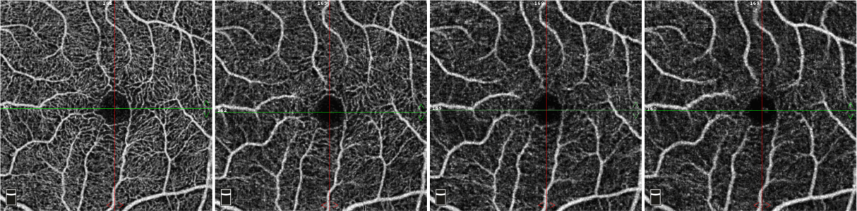
Common OCT angiography (OCTA) artifacts have previously been extensively described. The consequences of defocus have been shown, but the quantitative significance of even subtle changes in defocus have not been previously reported. After receiving institutional review board approval, sequential OCTA scans (AngioVue, Optovue, Fremont, CA) of a single eye of a normal participant were obtained; the scans demonstrated a purposeful progressive increase in the manual defocus setting from the optimum (Fig 1, A-D). Incorrect focus settings limited the ability to visualize fine capillary vessels and caused an apparent thickening of the remaining vasculature. Consequently, vessel density outputs in the retina decreased steadily (56.6%, 50.1%, 48.1%, 46.2%, Fig 1, EeH, respectively). Despite this, the signal strength index remained above the accepted quality threshold of 55 for all images. Therefore, when assessing an OCTA image without obvious motion artifacts, signal strength index is necessary, but not sufficient, for high-quality OCTA scans and valid data. Attention to image focus is paramount, and the presence of defocus of the retinal vasculature in OCTA datasets may elude casual inspection and result in inaccurate quantification.